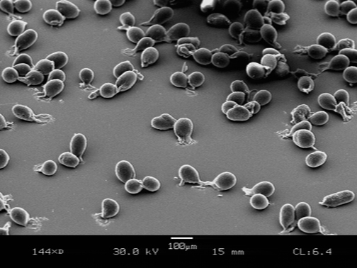
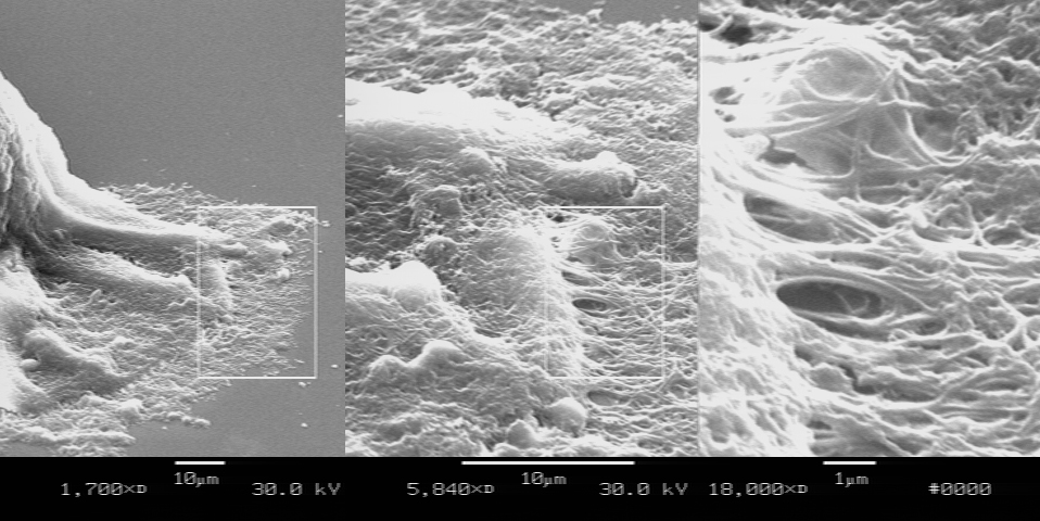
We know remarkably little about the structure and form of algal gametes within the Bryopsidales
Here are a few images of the large macrogametes typical of species of Penicillus, Rhypocephalus, and most Udotea

The SEM images of Rhipocepalus phoenix macrogametes shown here were obtained with assistance from Dr Ed Florance at Lewis and Clark College


